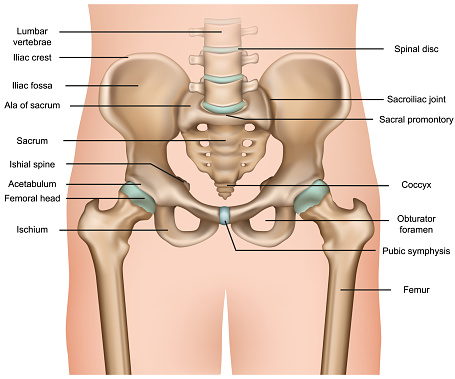
X-ray of a woman's pelvis. The joint gap between the ball and socket of the hip joint is reduced, partially or nonexistent. Where the bones of the joint meet, the surrounding cartilage has uneven edges and appears to be frayed. This abrasion is usually considered to be a sign of wear and tear or age.
Foto de Hueso de la cadera de la pelvis femenina humana, imagen de rayos X. De cerca
Imagen Disponible en Alta Resolución, Descarga inmediata
small: 680 x 518 Pixels, medium: 1177 x 896 Pixels, large: 1987 x 1513 Pixels, x_large: 4310 x 3280 Pixels,


